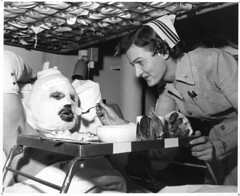
U.S. strategy for treating troops wounded in Afghanistan, Iraq: Keep them moving
By David Brown
Washington Post Staff Writer
Saturday, November 27, 2010
http://www.washingtonpost.com/wp-dyn/content/article/2010/11/27/AR2010112702875.html
An unofficial blog about the National Museum of Health and Medicine (nee the Army Medical Museum) in Silver Spring, MD. Visit for news about the museum, new projects, musing on the history of medicine and neat pictures.
U.S. strategy for treating troops wounded in Afghanistan, Iraq: Keep them moving
By David Brown
Washington Post Staff Writer
Saturday, November 27, 2010
http://www.washingtonpost.com/wp-dyn/content/article/2010/11/27/AR2010112702875.html
-an NPR followup -
by NPR Weekend Edition Sunday November 28, 2010
http://www.npr.org/2010/11/28/131644641/92-years-later-a-sickle-cell-surprise
-and finally the original short report –
Sheng Z-M, Chertow DS, Morens D, Taubenberger J. Fatal 1918 pneumonia case complicated by erythrocyte sickling [letter]. Emerg Infect Dis. 2010 Dec; http://www.cdc.gov/eid/content/16/12/pdfs/10-1376.pdf


Radiation Worries for Children in Dentists’ Chairs
November 22, 2010
http://www.nytimes.com/2010/11/23/us/23scan.html
The "Contributed Photographs" collection, as it came to be known, consists of photographs donated or contributed to the Museum. Photographs arriving during and after the war were usually added to the Surgical Section and numbered like the bones were. Many photographs were sent by doctors who wished to see their cases included in the History. Doctors such as Reed Bontecou of Harewood Hospital in Washington, J.C. McKee of Lincoln General Hospital in Washington (who also provided surplus photographic equipment after the Museum's burglary), and J.H. Armsby of Ira Harris General Hospital in Albany, New York, contributed dozens of photographs at the end of the war. They received photographs from the Museum in exchange. Most of the photographs given to the Museum were albumen prints, but infrequently a tintype (a photograph printed on thin metal) was donated. (Otis to Lyster, May 11, 1866) Tintypes were never as popular as other photographs. (Welling, p. 117) Their dark background made medical subjects harder to see and reproduce in print.
Otis frequently wrote to surgeons requesting a photograph of a specific case which he would then have engraved for the History. He also wrote to patients asking them to have their wound photographed. Otis wrote to Charles Lapham, who had been with Co. K of the 1st Vermont Cavalry:
The interesting report of your case, which is recorded
in this office, leads me to desire to possess if possible, a
photograph which shall farther illustrate it. The Surgeon
General possesses photographs of a number of the very rare
cases in which patients have survived after the very grave
mutilation of the removal of both thighs, and has instructed
me to request you to have a photograph prepared, the expense
to be defrayed by this office.
It would be well to have two pictures taken: one
representing the stumps, the other the appearance with
artificial limbs attached.
The photographer might take two or three prints of each
to be retained by you, and then should forward the
negatives, carefully packed to this office, by express,
enclosing at the same time the bill for his services.
I enclose copies of a photograph of the size desired.
(Otis to Lapham, May 25, 1865)
Lapham had the work done and two photographs were added to the collection.
Otis commissioned physicians such as E.D. Hudson of New York City to take photographs for him. Writing to Hudson, Otis said "I am anxious to obtain photographs of double amputations of the thigh or leg and of other cases of unusual interest, and am willing to pay for such. I hereby authorize you to have photographs taken of cases of especial interest. As near as may be they should be uniform in size with those taken at the Army Medical Museum, of some of which you have copies." In the same letter, Otis sent a list of soldiers who had survived the operation of the excision of their humerus. Hudson, a maker of prosthetics, undoubtedly appreciated Otis' fulfilling his request for the names. Otis and Hudson's arrangements to look out for each others interests, resulted in striking photographs such as the two of Columbus Rush, a young Confederate from Georgia who lost both legs. (Otis to Hudson, February 7, 1866) Otis and Hudson cooperated so closely that Hudson was able to display his prosthetics in the Medical Department's exhibit at the Centennial fair. (Otis to Hudson, March 8, 1876)
For many years, these photographs received a Surgical Section number and were bound in volumes labeled Photographs of Surgical Cases. (Otis to Washburne, April 4, 1866) The photographs donated to the Museum were often rephototographed to be included in the Surgical Photograph series. Roland Ward's plastic surgery after the destruction of his lower jaw (SP 167-170, 186) is an example. Columbus Rush's photograph, in which he demonstrates his Hudson-made artificial legs, was copied and sent out as part of the series. Otis also purchased photographs from studios, buying "two dozen of the war views for the Museum" from E. & H.T. Anthony & Co. (Otis to Anthony, September 25, 1865)
Contributors of photographs like Hudson also used the pictures themselves. Dr. Gurdon Buck is particularly noteworthy for his use of photographs. He had engravings made of "before and after" photographs for his 1876 text on plastic surgery, Contributions to Reparative Surgery. In the engravings, Buck used drawn lines to explain his operation. Buck deposited a set of his photographs in the Army Medical Museum soon after the end of the war.
About 1876, as photographs of many sizes and from many people continued to arrive, the collection was removed from the Surgical Section and named the Contributed Photographs. Otis no longer had the photographs bound in albums. All of the photographs were renumbered from the beginning in red ink with the identifying "Cont. Photo." or the initials "C.P."6 Some of the best photographs were copied in the Museum and published as part of the Surgical Photograph series. Others were engraved for the History. Some photographs almost certainly taken by the Museum such as the one of Neil Wicks, probably by Bell,7 were added to the collection after the original negatives disappeared. Unfortunately, many photographs were given away by Daniel Lamb in 1915 including scores to Reed Bontecou's son.

Curatorial Records: Numbered Correspondence 317
November 19, 1894
Dr. Judson Daland
319 S. 18th St.
Philadelphia, Pa.
My Dear Doctor:
Can you give me any information concerning a centrifugal machine which is considered superior to the Litten centrifugal? Dr. Gray has just informed me that you are using a superior machine for urinary and blood analysis, and hence I write to ask you that you will be kind enough to enlighten me on this subject, especially as Surgeon General Sternberg is considering the matter of supplying certain of the larger military posts with the latest and most improved centrifugal apparatus.
Very sincerely yours,
Walter Reed
Major and Surgeon, U.S. Army,
Curator Army Medical Museum.
Curatorial Records: Numbered Correspondence 7132
November 18, 1903.
Mr. Henry Reens,
409 Fourth Ave.,
New York, N.Y.
Dear Sir:
In accordance with your request of the 17th inst. 6 copies of printed circular of Museum photograph 177, recovery after fracture of the right ilium by a musket ball (from your own case), are herewith forwarded.
Very respectfully,
C.L. Heizmann
Col. Asst. Surgeon General, U.S.A.
In charge of Museum & Library Division