Today Kathleen and I joined the staff of the Borden Institute (who publish the Textbooks of Military Medicine) to keep working on a Walter Reed Army Medical Center Centennial Atlas, ie a book of photographs of 100 years of it being a hospital. We're going to make a big push in January to finish the book which should be available in late April. Watch this space.
In the meantime, we still need photographs of the base from the 1970s-1990s. If you were at Walter Reed and have pictures, let us know.
Friday, December 5, 2008
Who writes this stuff anyway?
Not enough of us, that's who. It's mainly the Archives staff, so I removed all the people who never have posted to the site. That's why the list of names in the upper right corner suddenly shrunk.
Sadly, some losses
Today the AFIP director reported, "Dr. Ahmed Hidayat, Chief Ophthalmic Pathology, AFIP, passed away last evening from a long-term illness. Dr. Hidayat was a long-time member of the Institute Staff in Ophthalmic Pathology."
And STIL Casing Solutions (whom we bought 16mm film cans from for our eventual film project) sent an email telling me that André Pion, the person who I usually dealt with and just talked to a couple of weeks ago about new DVD cases, passed away too.
We regret to announce the death of our colleague and friend André Pion, who passed away last Tuesday evening from septicaemia (blood poisoning). Death’s irrevocable nature makes it very difficult to accept, but at the same time reminds us of how priceless life is.
We will remember him for his unquestioned integrity, intellectual honesty and his devotion to his work, and also for the gifts of his friendship and humour. He cared deeply for each person he talked with, he loved his work and felt privileged to be able to do something he loved every day.
Thursday, December 4, 2008

Surgical Photographs
I've been reviewing the work our scanning contractor has been doing for us. It's a never-ending job because of the volume of images they're handling. We actually scanned the Surgical Photos in-house and sent them and the database to the contractor for upload, but I'm still going through them to make sure we sent all versions of a particular case. For instance, many of the photos in this collection are of Civil War soldiers showing their healed wounds, and many of those are wounds or amputations of the leg up to the hip. These men were often photographed without draping them in some way to protect their modesty. I personally am surprised at that, but that's how it was done.
However, some of these photos were displayed at the 1876 World Exposition in Philadelphia and it was then that modesty prevailed. Or, rather, as Mike and J.T.H. Connor wrote in Shooting Soldiers: Civil War Medical Images, Memory, and Identity in America, it appeared that the issue was less about protecting the men's identity and modesty than it was about not offending the potential audience.
In any case, we have more than one version of some of these photos: those with fig leaves and those without, and I've been going through the 400 in the collection to make sure that all versions were uploaded.
Not all of the photos are of soldiers, though. Here's one of a young boy who was shot in the head with a shotgun. It's called Successful Operation of Trephining of Cranium for Gunshot Injury.

And here's the case history:

Wednesday, December 3, 2008
A couple of pictures
Nothing special about these. They just appealed to me.
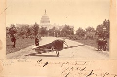
CP4355, Travois Litter [in front of Capitol building, Washington, DC] by Capt JC McDonald, photograph by CM Bell.
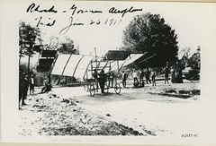
Reeve 072203, Airplane, 1910. "Rhodes - Gosman aeroplane trial, 01/26/1910. Fort Barrancas, Florida."
CP4355, Travois Litter [in front of Capitol building, Washington, DC] by Capt JC McDonald, photograph by CM Bell.
Reeve 072203, Airplane, 1910. "Rhodes - Gosman aeroplane trial, 01/26/1910. Fort Barrancas, Florida."
New exhibit
The Historical Collections guys and the exhibit guy finished putting together an exhibit yesterday and I went over and shot some of the process as well as the finished product. Because I know for a fact, yes a fact, that not one of them will write about it, I'm doing it because I'm so responsible. And because I love behind-the-scenes stuff and assume you do too.
The exhibit is contained in one wall-mounted cabinet and is called Facial Reconstruction. We have really cool and interesting plaster models and they're what make up the bulk of the cabinet. Here are four on them on a cart, waiting to go into the cabinet. They're various stages of one person's reconstruction.

Here are two of the three guys working on the cabinet.

They used the line of the bottom row of models (the ones shown on a cart above) to mark a line for the next row up. Here's that bottom row being hung.

Here's the exhibits guy using a spiffy, bendy thing on the drill to make a hole for the next row up.

A test fit on the second row.

Here's a close-up of them on a cart.

The models are all safely tucked away again and the labels are installed.

Here are a couple different models, both from World War 1. The first one shows a nasal splint after the surgeon rebuilt his nose from a flap of skin from his forehead. Note the scar.

This one shows an appliance used to keep his fractured upper jaw aligned correctly within his face.

This is a more contemporary model. This man sustained a severe head injury and a portion of his skull was removed to allow his swollen brain to expand. A CT scan of his head allowed the doctors to create a resin model of his skull and then make a cranial plate based on a mirror image of the undamaged side of his skull. This view shows a portion of the skull removed. It's art, isn't it?

And finally, the finished exhibit. Ta-Da!!

Tuesday, December 2, 2008
Browsing
Today we had a request for images of people who were blinded by poisonous gas. If the requester had asked for rabbits we would have been in business, but we had nada for those two conditions together. Some blindness, some poisonous gas, but the Venn diagram did not converge.
I did find, however, some interesting pictures about blindness, and here they are.
Reeve 870, A blinded French soldier, World War 1

Reeve 871, A blinded French soldier and his bride, World War 1
![]()
AEF007 (American Expeditionary Forces)
Blind French soldiers, patients in the department organized by Miss Winnifred Hope for the re-education of the blind. Base Hospital number 115, Hotel Ruhl. Base Laboratory Hospital Center Vichy, France. 08/1918[?].

Reeve 14494: American Red Cross workrooms. Paris, Seine, France. Stitching eye bandages on the machine in the American Red Cross workrooms for surgical dressings, rue de la Faisanderie, Paris. These bandages are used largely for gas cases.

I did find, however, some interesting pictures about blindness, and here they are.
Reeve 870, A blinded French soldier, World War 1

Reeve 871, A blinded French soldier and his bride, World War 1

AEF007 (American Expeditionary Forces)
Blind French soldiers, patients in the department organized by Miss Winnifred Hope for the re-education of the blind. Base Hospital number 115, Hotel Ruhl. Base Laboratory Hospital Center Vichy, France. 08/1918[?].

Reeve 14494: American Red Cross workrooms. Paris, Seine, France. Stitching eye bandages on the machine in the American Red Cross workrooms for surgical dressings, rue de la Faisanderie, Paris. These bandages are used largely for gas cases.

Tuesday, November 25, 2008
Audio Tour at the NMHM
About a year ago, the Museum acquired the Tour-Mate audio tour system, which allows visitors to do a self-guided highlights tour of the permanent exhibitions. Just yesterday, we added an additional hour to the audio tour to include the new exhibit "RESOLVED: Advances in Forensic Identification of U.S. War Dead," and “Trauma Bay II, Balad, Iraq.” Come by the museum for a listen. We might even have the files available for download on our website soon.
St Elizabeths hospital history
We've got a lot of autopsy records from St Elizabeths hospital in our Neuroanatomical collections. A new article discusses the race relations at the hospital, especially between the long-term patients and the soldiers arriving after WW1. Ask for an interlibrary loan of "`These strangers within our gates': race, psychiatry and mental illness among black Americans at St Elizabeths Hospital in Washington, DC, 1900-40" by Matthew Gambino, History of Psychiatry, 19:4, 2008. I read it at work today - Matthew's used our collection in the past although not for this article.
Monday, November 24, 2008
Telemedicine from the first world
Here's an Washington Post article about a British couple who have set up their own charity to provide telemedicine around the world, based on just themselves, an assistant and a lot of energy. The Swinfen Charitable Trust sounds like a pretty amazing shoe-string operation. Based in England, it has links to the University of Virginia. It's apparent in this article that telemedicine is going to change the practice of medicine as the 21st century progresses.
New upload to the Internet Archive
Today we uploaded a new item to the Internet Archive. It's "A Guide for Uniform Industrial Hygiene Codes or Regulations for the Use of Fluoroscopic Shoe Fitting Devices," by The American Conference of Governmental Industrial Hygienists.
It sounds kind of boring. All right, it sounds really boring, but when you read it you have to say to yourself, "what were they thinking?" It's self-described as a guide "designed to minimize the amount of radiation to which persons are exposed during the use of fluoroscopic shoe fitting devices." In other words, shoe stores had x-ray machines that you stuck your feet in (and our museum has one of them (the machine, not the feet)) to see how well your shoes fit. I dunno, when I was a kid the salesman used to press down on the toe of the new prospective shoes and ask if I could feel it.
Anyway, you can see this guide here.
Blackhawk as sickbed reading, circa 1951

Here's a picture that one of the assistant archivists brought to my attention today. This poor guy has a gunshot wound of his lower femur (shown with a Blackhawk comic book on the bed) during the Korean War, 1951.
Scanned on a computer old enough to require a scuzzy port to connect to the scanner, copied to a cd and then carried home to be uploaded to Flickr and blogged about.
Saturday, November 22, 2008
Photos aren't us continued again
Thomas asked "what's going on" in a previous posts comments. I have no idea why Flickr is blocked. However for the USB ports, this is a response to a computer virus - kind of like using a sledgehammer to swat a fly.
Oddly enough, Australian papers rather than American ones seem to have picked the story up and here's one. This earlier Wired article says:
The problem, according to a second Army e-mail, was prompted by a "virus called Agent.btz." That's a variation of the "SillyFDC" worm, which spreads by copying itself to thumb drives and the like. When that drive or disk is plugged into a second computer, the worm replicates itself again — this time on the PC. "From there, it automatically downloads code from another location. And that code could be pretty much anything," says Ryan Olson, director of rapid response for the iDefense computer security firm. SillyFDC has been around, in various forms, since July 2005. Worms that use a similar method of infection go back even further — to the early '90s. "But at that time they relied on infecting floppy disks rather than USB drives," Olson adds.
So this is a problem that dates back 2 decades and was apparently addressed by anti-viruses, but this is the current response. Personally I think there's a second underlying reason and this virus is just the current cover story. However, USB ports and the Internet are the way computers work now - as much as the military would like to, they're not going to be able to singlehandedly reset technology to 1995 nor return the Internet to a DARPAnet.
I put in a request to have my scanner port opened again, but I honestly do not expect to get a response. At some point, probably right about now, having computers on the military's network will be too much trouble and I'll pull them all to stand alone. People can just go back to telephoning with their requests - which we will then be able to actually fulfill.
Friday, November 21, 2008
More discoveries
I found this series when doing research for someone the other day.
The initial photo of Albert Bauer, a soldier wounded in World War 1:

The first medical illustration demonstrating the surgical procedure used to correct it:

And the continuation of the procedure:

I haven't come across the final picture but hope I do. I'd really like to see the finished reconstruction.
Osler photos
And, like yesterday, here's an announcement of someone else's neat history of medicine website. At one point in the early 20th century, the Museum rebuilt McGill's medical collections after a fire. One of their professors has rediscovered what's left recently, and I'll try to post on that soon. In the meantime, check this out:
The William Osler Photo Collection
The McGill Library is pleased to launch The William Osler Photo Collection, a searchable and browsable website of 384 images drawn from the Osler Library’s collection of photographs of Sir William Osler (1849-1919), who graduated from Medicine at McGill University in 1872 and, after a brief interval, taught there for ten years. He went on to the University of Pennsylvania (1884-1889), Johns Hopkins (1889-1905) and finally became Regius Professor of Medicine at Oxford and one of the most famous doctors in his time. There are photographs from all stages of his life, along with pictures of Lady Osler, his son Edward Revere Osler and other family members. The site was made possible by a generous donation from the John P. McGovern Foundation.
The url is http://digital.library.mcgill.ca/osler/
Photos aren't us continued
Today the AFIP's IT department reached in and turned off our USB ports so we no longer have access to the 3/4 of a terabyte of hi-resolution scans on our external harddrives. They also made our scanners non-functional at the same time, as they plug into USB ports, so we can't make new scans for people either.
On the positive side, I talked with an ex-AFIP staffer who worked in the Medical Illustration Service from the early 1960s through the mid-1970s. He's given Historical Collections a moulage kit he worked on and we're going to do an oral history with him.
On the positive side, I talked with an ex-AFIP staffer who worked in the Medical Illustration Service from the early 1960s through the mid-1970s. He's given Historical Collections a moulage kit he worked on and we're going to do an oral history with him.
Thursday, November 20, 2008
In other news... excellent History of Embryology site launches

This press release came through the Caduceus history of medicine list today:
Making Visible Embryos, http://www.hps.cam.ac.uk/visibleembryos/
An online exhibition by Tatjana Buklijas and Nick Hopwood, Department of History and Philosophy of Science, University of Cambridge, with funding from the Wellcome Trust.
Images of human embryos are everywhere today: in newspapers, clinics, classrooms, laboratories, baby albums and on the internet. Debates about abortion, evolution, assisted conception and stem cells have made these representations controversial, but they are also routine. We tend to take them for granted. Yet 250 years ago human development was nowhere to be seen.
This online exhibition is about how embryo images were produced and made to represent some of the most potent biomedical objects and subjects of our time. It contextualizes such icons as Ernst Haeckel's allegedly forged Darwinist grids and Lennart Nilsson's 'drama of life before birth' on a 1965 cover of Life magazine. It also interprets over 120 now little-known drawings, engravings, woodcuts, paintings, wax models, X-rays and ultrasound scans from the fifteenth to the twenty-first century. It displays the work of making visible embryos.
Contact: hps-embryo@lists.cam.ac.uk

One image on their site is from our museum - a His Embryograph - but we have similar collections of wax models, embryos and embryo models as discussed in the article. The two photographs here are from our collection. Some of the embryological collection is on display and I've heard that a reworking of it is underway.
Photos aren't us
As you've seen previously, last week Walter Reed blocked access to Flickr so we can't upload photographs for people to use or just enjoy. At the same time, they also blocked access to uploading services such as Rapidshare or Yousendit that we had been using to send photographs to requestors with same-day service. We switched back to burning and mailing cds this week. However today, the military implemented a policy of blocking USB ports on all networked computers (see below for details), and since they had previously required all their computer networks to be hooked together (changing our email addresses overnight but not actually notifying us about the change so all our email was bouncing), we're affected . Since all of our gigabytes of hi-resolution scans are on external hard drives that connect via USB, and we can't upload pictures to the internet, we are at the moment out of the photo library business and will not be providing publishable quality images to researchers. We may still be able to email small images. We apologize to our users. To be honest, since the CAC cards required to turn on the computers, the mouse and keyboard are all via USB, I don't actually expect to have a functioning computer at work. I would suggest calling the Museum if you have a question about coming in to do photo research since we will still be able to provide you access to the original image, unless it was electronic in the first place.
Here's the policy as sent out by Walter Reed's Department of Information Management (DOIM):
Effective immediately, the use of USB storage devices are suspended on all DoD NIPRNET and SIPRNET computers.
This rule will be technically implemented beginning 19 1800 November 2008 and will be applied across the entire network on all computers. Implementation of this rule will impact all memory sticks, thumb drives, USB external hard drives, and camera flash memory cards. USB connected printers with internal and external media storage (e.g. SD Cards, etc.) may also be impacted.
Other USB connected devices such as keyboards, mice, CAC readers, and blackberries "SHOULD NOT" be affected. Any user that experiences problems with such devices after technical implementation is asked to call the DOIM help desk or follow the procedures noted below for faster service.
These actions are being completed as part of an Army-Wide Information Assurance initiative to protect the DOD network from intrusion and continuous attacks. In order to further protect our network we ask all users to adhere to posted rules and allow us the opportunity to find secure alternatives (if those exists) to meet mission needs.
Wednesday, November 19, 2008
Today's discoveries
Did I ever tell you how much I like my job? Sometimes there's too much of it, but usually it's a journey of interesting discoveries.
Today I worked on a reference request that included anything we have on the Polish Relief Commission in World War 1. Searching for images for someone else is almost like spending someone else's money. I have all the fun and it's on someone else's dime. Cool.
We have about 100 pictures that include the Commission's name in the caption. Some of them, like some of just about everything, are, sorry, boring, but some just grab you right off and demand a second look.
Here's what I mean.

Reeve 31754 Polish Relief Commission (Col. Gilchrist). Little Polish girl.

Reeve 31756 Polish Relief Commission (Col. Gilchrist), opening public bathing place, distribution of cigarettes.

Reeve 31765 Polish Relief Commission (Col. Gilchrist), three waifs, fatherless & motherless, from effects of typhus fever, near Dora-Husk, Poland, 1920.

Reeve 31770 Polish Relief Commission (Col. Gilchrist), delousing Bolsheviks on the highways, 1919-1920.

Reeve 31933 Polish Relief Commission. (Col. Gilchrist). American equipment in the near east. Foden Thresh steam sterilizer with A.P.R.E. to Poland.

Reeve 31935 Polish Relief Commission (Col. Gilchrist). Cases of cholera left by retreating Bolsheviks near Villna, 1919.
Now admit it. Don't I have a great job?
Subscribe to:
Posts (Atom)