I was tickled pink to find this reply to the doctor who sent his wax preparations to the Museum (letter published March 15) and who said it would break his heart if the liver were broken.
April 1, 1887.
Dear Doctor:
Your note of March 30th is received. I am very sorry to say that the preparation of the liver was smashed into powder. But as it had been delivered to Dr. Wortman I felt bound to pay for it, although it was not possible for me to certify that it had been received, and the only thing I could do was to pay for the lung. The risks of transportation of such specimens are evidently much greater than I had supposed, and I do not think I will try it again. Some day I hope we can make such preparations here. Dr. Wortman will write you explaining how it happened. He had a pleasant visit and acquired much valuable information. Accept my sincere thanks for the very courteous manner in which you received him. With best wishes believe me to be
Yours very sincerely
(Signed) John S. Billings.
Dwight Prof. Thomas
Harvard Medical School,
Boston, Mass.
P.S. The injection of the kidney has come to hand in perfect condition.
Showing posts with label wax models. Show all posts
Showing posts with label wax models. Show all posts
Thursday, April 1, 2010
Monday, March 15, 2010
Letter of the day, March 15
Harvard University
Medical School
Anatomical Department
Boston, March 15, 1887
My dear Dr. Billings,
I have made three more corrosion preparations for your museum. The first is a left human lung [A.M.M. No. 2432 Anatomical Sect.] – vein red, artery blue. It is decidedly better than the one that was broken and which it is sent to replace. It shows the shape of the lung including the curve cut out to make room for the heart. There is an extravascetion[?] at one place but it does not show very much. I have made also a human liver in four colors. Portal vein red, hepatic vein (and cava) blue, artery yellow, bile duct green. It is the best preparation of the kind that I ever made. The only defects are that the acid has affected the green which has unfortunately become very bluish and that the yellow [section of page torn off] I think the preparation [torn] called one of the first class. The kidney which I sent [illegible] had rather a weak injection of the vein. This is perhaps as well as it shows more of the rest but I have now a preparation which is its complement.[A.M.M. No. 2434] Namely a full injection of the vein in blue, the ureter in yellow and no artery.
The lung is sent to replace the other one. The price of the liver is fifty dollars and the kidney is thrown in. The lung and liver are mounted elastically on cushions covered in white silk. I hope you will send a man for them as it would break my heart if the liver were broken.
I intend now to give up corrosions. They take too much time and should be made by demonstrators not professors. A visitor just came in to see the liver. I think it has changed shape a little by its weight. It is worth sending for anyway and if it deteriorates you can pay what you please.
Yours very sincerely,
Thomas Dwight
[Specimens of lung & liver received Mar. 28, 1887
Kidney Apr 1 ‘87
Liver broken when received and not placed in A.M.M. Could not be repaired.]
Thursday, March 4, 2010
Letter of the day, March 4
We still have many wax models, showing just the kinds of things he's asking for in this letter.
GRC/mj
War Department
Office of the Surgeon General
Army Medical Museum and Library
Washington
March 4th, 1919
Circular Letter No. 121.
Subject: Reproduction of Interesting Lesions in Wax.
1. There is present at the Army Medical Museum an expert in the reproduction of various lesion of the skin in wax. A considerable number of models have been made during the war and it is desired to make this collection as excellent and as representative as possible.
2. The following types of cases can be well represented in wax: chronic or unhealed ulcers following various types of wounds; unhealed lesions resulting from gas burns; unusual scar formations; and unusual skin diseases. Such lesions can be most naturally reproduced by wax models and it is believed that many of the hospitals receiving cases from overseas have cases of this nature which should be reproduced for permanent record.
3. As it is impossible for the one worker in wax models to travel from place to place, it is requested that when such cases occur at any Army hospital they be reported to the Surgeon General’s office, attention the Laboratory Division, with a brief description of the case and probable permanence of the lesion at the time, accompanied by a rough unmounted photograph if possible to obtain the same.
4. It is intended to order especially interesting cases of this character to the Walter Reed Hospital for further treatment and for the production of the model which will be a permanent exhibit in the Army Medical Museum.
By direction of The Surgeon General:
C.R. Darnell,
Colonel, Medical Corps, U.S.A.
Executive Officer
Copy to:
Commanding Officers of all
Base Hospitals,
General Hospitals,
Embarkation Hospitals
Saturday, February 13, 2010
Letter of the Day: February 13
111 Bruce Ave
Yonkers N.Y. Feb. 13th 1904
Surgeon General R.M. O’Reilly, U.S. Army
War Department, Washington D.C.
Dear General:
During the last thirty years I have made a collection of anatomical and pathological material consisting mostly of wax models in colors illustrating deformities of the nose, mouth, throat and chest. These have been made from casts taken from the subjects before and after operation.
The above collection I am considering presenting to the Army Medical Museum in case the museum would be pleased to receive the same.
Some months ago I was contemplating a visit to the museum when I hoped to have the pleasure of meeting you. Owing to illness my condition will not allow of it I will enclose a note of introduction from my friend Dr. J.S. Billings.
I will appreciate it if you will kindly advise me regarding the reception of the collection and the facilities you have for exhibiting the same. I will be pleased to give you detail information of the collection should you desire it.
Awaiting your reply, I am
Very respectfully yours
D.H. Goodwillie M.D.
per R
Dictated by Dr. Goodwillie
[A five-page list of models was in the file with the letter.]
Tuesday, July 29, 2008
Let's look at a photo
This one's for Johanna of the Morbid Anatomy blog - she's seen this picture before, but let's take a look at it.
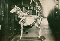
Comparative anatomy, Auzoux model of horse, life size. Specimen no. 2635. [papier mache, on display in Army Medical Museum]. We no longer have the model, although one can be seen in the Science Museum in London.
For many years, models were a way to convey information in medicine and natural sciences. Color printing had to be hand-done, and photography first didn't exist, and then each photograph for a book had to be printed individually and glued into the book. And hand-tinted if required. So well into the 20th century models like Auzoux's above, or ones like this x-ray burn were produced for education.
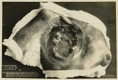
Breast. Burn, X-Ray. Wax model. No. 92 X-ray burn involving right breast and axilla. Necrosis of tissue producing sloughing ulceration, the bottom of which includes pleura and lung tissue. X-ray treatment was applied for carcinoma of the breast. Colored, woman, age 35 years. Army Medical Museum model prepared by Dr. J.F. Wallis. [Circa World War 1, 1918].
The museum's model-making skills continued into the 1950s, when a technique called moulage, which simulated injuries in rubber overlays were developed. A soldier would put on a moulage of a nuclear radiation injury for example and then the trainees would attempt to treat him. I'll attempt to get some photographs of them up, although I think we scanned the kit's whole instruction book recently as well.
Check out this conference in Europe on models and Auzoux too.
Comparative anatomy, Auzoux model of horse, life size. Specimen no. 2635. [papier mache, on display in Army Medical Museum]. We no longer have the model, although one can be seen in the Science Museum in London.
For many years, models were a way to convey information in medicine and natural sciences. Color printing had to be hand-done, and photography first didn't exist, and then each photograph for a book had to be printed individually and glued into the book. And hand-tinted if required. So well into the 20th century models like Auzoux's above, or ones like this x-ray burn were produced for education.
Breast. Burn, X-Ray. Wax model. No. 92 X-ray burn involving right breast and axilla. Necrosis of tissue producing sloughing ulceration, the bottom of which includes pleura and lung tissue. X-ray treatment was applied for carcinoma of the breast. Colored, woman, age 35 years. Army Medical Museum model prepared by Dr. J.F. Wallis. [Circa World War 1, 1918].
The museum's model-making skills continued into the 1950s, when a technique called moulage, which simulated injuries in rubber overlays were developed. A soldier would put on a moulage of a nuclear radiation injury for example and then the trainees would attempt to treat him. I'll attempt to get some photographs of them up, although I think we scanned the kit's whole instruction book recently as well.
Check out this conference in Europe on models and Auzoux too.
Wednesday, May 21, 2008
A day in the life...
An interesting research request came in through the Radiology department today. Someone's looking for fluoroscope burns. So far I haven't turned up exactly what they want, which is modern color shots, but check out this photo of a wax model that I did find:
The caption reads: Breast. Burn, X-Ray. Wax model. No. 92 X-ray burn involving right breast and axilla. Necrosis of tissue producing sloughing ulceration, the bottom of which includes pleura and lung tissue. X-ray treatment was applied for carcinoma of the breast. Colored, woman, age 35 years. Army Medical Museum model prepared by Dr. J.F. Wallis.
Wallis means that it was done during World War 1, because that's when he was on the staff. We might still have this model in historical collections, but that department was working at the warehouse today, and thus missed the coffee and cake that we had to celebrate the Museum's birthday.
So how does one find something in the Archives? We've got an internal database (or fifty) that you can access a derivative of at our Guide to Collections. With the help of contract Archivists from the Information Manufacturing Company, we're scanning tens of thousands of images per year and uploading them into an internal database, only available to our staff now, but eventually we'll open it to a wider audience. And some of the finding something is me or one of the other archivists knowing where something is because we put it away a decade ago or so.
Saturday, May 10, 2008
Anatomical Theatre website launches
Morbid Anatomy's launched a new site based on an exhibit of photographs she's done. She writes "I have finally launched the website for Anatomical Theatre, the photographic exhibition of medical museum artifacts. For more information about the project, check out the "Introduction" and "Press Release" pages."
Wax and plaster models as well as other specimens from the NMHM are included.
Wax and plaster models as well as other specimens from the NMHM are included.
Subscribe to:
Posts (Atom)